

Birth
In a second-floor tenement flat at 10 Duntroon Street. 1930
Education
Whitehill Senior Secondary School, Dennistoun, Glasgow. 2007
Death
At 56 Sea Road, Bexhill-on-Sea, Sussex, England (from a heart attack).
Religion
AgnosticDr June Dalziel Almeida was an internationally renowned Scottish virologist. She pioneered new methods for viral imaging and diagnosis.
Personal Information
Name(s)
Almeida (née Hart) June Dalziel
Date and place of birth
5 October 1930, in a second-floor tenement flat at 10 Duntroon Street, Glasgow, Scotland.
Death and place of death
1 December 2007 at 56 Sea Road, Bexhill-on-Sea, Sussex, England (from a heart attack)
Family
Mother: Jane Dalziel (née Steven), shop assistant, born in Glasgow.
Father: Harry Leonard Hart, bus driver, born in London.
Brother: Harry Hart, died 1940, aged six years, from diphtheria [1].
Marriage and Family Life
(Mention women relatives by name and dates, especially if they are distinguished. Spouse and children.)
In 1954 June Hart married Henry (Enrique) Rosalio Almeida (1913‒1993), a Venezuelan artist. They had one daughter, Joyce (1960‒), born in Toronto, Canada.
In 1967 the Almeidas divorced.
In 1982 June Almeida married virologist Professor Phillip S Gardner (1925‒1994), followed by retirement in 1985 to Bexhill-on-Sea [1].
Education
June Hart flourished academically and received a high-quality education at Whitehill Senior Secondary School, Dennistoun, Glasgow. Her subjects included Latin and Greek, and in her fifth year she won a school prize in Science. June Hart played hockey, enjoyed ballet and in her teenage years developed a passion for photography [1].
Financial constraints prevented June Hart from going to university. Having passed her “Higher” examinations, she left school in 1947, aged 16 years.
In 1970 June Almeida was awarded a Master of Philosophy (MPhil) degree from the University of London; in 1971 she was awarded a Doctor of Science (DSc) from the University of London [2].
Religion
June Almeida described herself as an agnostic and always looked to science for explanations of life’s realities [3].
less
Significance
Works/Agency
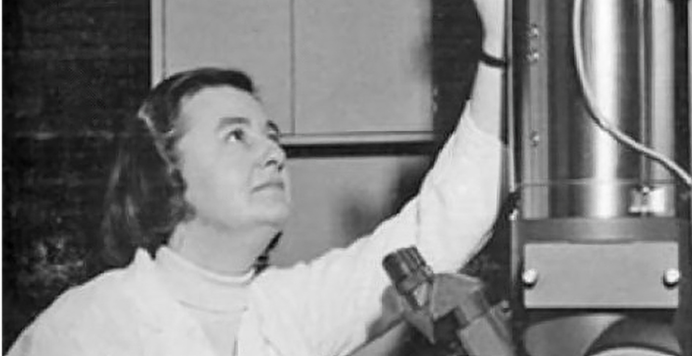
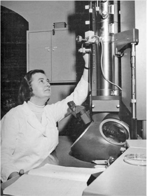
June Almeida was intensely creative, talented in photography (from her teenage years) and art (shown by her recognised skills in later life in china restoration). She felt that her ability to find and photograph viruses with the electron microscope (EM) was inherently linked to her wish to find and take a great photo. She excelled at composition and recognizing patterns [1].
After leaving school, June Almeida’s decision to pursue a career in the biological sciences may have been influenced by the death of her younger brother from diphtheria [1].June Almeida had a high IQ and was a member of MENSA [4].
Reputation
(When alive, over time and modern scholarship. Subject’s status at death i.e., did she die with recognition or has that been posthumous? If possible, track her appearance and disappearance over time.)
With a starting salary of 25 shillings a week, June Hart began her working life as a junior histopathology technician at the Glasgow Royal Infirmary. In 1952 the family moved to London, where June Hart was appointed Research Assistant to Professor John WS Blacklock (1896‒1973) in the Pathology Department of St Bartholomew’s Hospital. In the same year June Hart became an Associate of the Institute of Medical Laboratory Technology [2], the forerunner of today’s Institute of Biomedical Science .
Marrying in 1954, the Almeidas emigrated to Canada in 1956,
In 1956 June Almeida’s work with the EM ‒ of which she had no previous working experience ‒ began with her appointment as Research Assistant to Dr A F Howatson in the Division of Biological Research, Ontario Cancer Institute (OCI), Toronto, Canada.
After the invention of the EM in 1931, following which the first commercial instrument became available in 1939 [5], electron microscopy was an emerging field of study in the 1950s. June Almeida’s aptitude with the EM was apparent, and her reputation grew. For example, her first peer-reviewed journal article [6] ‒ describing “a procedure that allows cells grown in vitro to be sectioned in a plane parallel to the glass surface to which they are attached” ‒ was submitted for publication only one year after starting work in Toronto.
Not having a university degree did not prevent June Almeida’s career from blossoming, and it has been suggested that in Canada and the North American continent the lack of such a qualification did not obstruct career progression compared to the United Kingdom [7]. Thus, between 1958 and 1983 June Almeida authored or co-authored 103 peer-reviewed papers or chapters and was lead author of 64 [2].
The technique of negative contrast staining was originally applied to purified virus preparations [8]. However, June Almeida refined this technique, enabling the use of crude preparations to study cell-associated viruses, and which offered “a new approach to the study of the morphology of basic cell components in the normal cell and the virus cell relationship in infected cells” [9]. Explaining the technique herself, June Almeida wrote: “The virus particle is surrounded by heavy metal atoms which act as an electron stain, the electron beam can pass through the low electron density of the virus but not through the metallic background, hence ‘negative stain’, a light virus against a dark background. Practically, the method is equally simple. A suspension of virus is mixed with a solution of the right kind of heavy metal ions, frequently those of phosphotungstic acid, and allowed to dry down onto a grid” [10].
Reflective of her approach to medical science, June Almeida’s EM work combined efficiency, focus and range. For example, within three years, she helped identify the morphological characteristics of wart viruses [11] and determined their sites of production [12]; members of this family ‒ later designated human papilloma viruses ‒ would be shown to cause cervical cancer, against which an effective vaccine is now available. June Almeida also described the morphology of the Herpesvirus varicella, making the important observation that “[t]he viruses of varicella and herpes simplex appear to be indistinguishable by morphological criteria …” [13]; and applied her evolving technical refinements to determine hitherto unknown structural aspects of rabies virus [14].
In 1963, June Almeida was promoted to Research Assistant at the OCI, and became a Fellow of the Royal Microscopical Society [2]. Meanwhile, she had been exploring the possibilities of immune-electron microscopy (IEM), to which she was introduced by an American visitor to the OCI [15].
IEM relies on the ability of a specific antibody to a specific viral antigen to clump together virus particles, which would otherwise be spread out and thus difficult to visualise. June Almeida’s insight was to realise that “IEM could help extend the range of what was possible with negative staining” [15] June Almeida and colleagues undertook initial IEM studies on wart and polyoma viruses [16] and “[t]o their delight the technique made smaller viral particles much more visible than previously thought” [15]. Crucially, IEM not only allowed negative staining to be used ‒ obviating the need for purification steps ‒ but also enabled viruses to be observed in situ and permitted the nature of antigen-antibody reactions to be investigated [15].
In 1964, Professor AP Waterson ‒ Chair of Microbiology at St Thomas’s Hospital, London ‒ visited June Almeida’s OCI laboratory and, impressed by her high-quality work, invited her to work in his department at St Thomas’s [7]. Professor Waterson and June Almeida made an ideal scientific combination, and Waterson ‒ unusually at that time ‒ unlike other professors of virology, was ahead of his time and saw that the clinical aspects of virology would flourish in due course [17].
Shortly after taking up her post at St Thomas’s as Scientific Assistant (funded by the Medical Research Council) [2], June Almeida made an important discovery, which decades later would bring her name to a wider audience beyond the field of virology.
Dr David AJ Tyrrell (1925‒2005), working at the Common Cold Unit, Salisbury, Wiltshire, England, had been investigating samples taken from pupils at a boy’s boarding school in the Epsom area of Surrey, England, one of which ‒ sample B814 ‒ could be passaged in organ culture and produce common cold symptoms in volunteers, but there were no unequivocal changes in the cultures [18]. In his memoir [19] Tyrrell recalls learning that June Almeida “was seemingly extending the range of the EM to new limits. It was generally believed at the time that you had to have concentrated and purified viruses in order to detect them with such an instrument. But she claimed that she would be able to find virus particles in our organ cultures with her new, improved techniques. We were not too hopeful but felt it was worth a try.” The sample was sent by train to London and June Almeida “saw virus particles in the B814 specimens!” [19].
She was confident that the B814 viruses were new, and after conferring with Tyrrell and Waterson ‒ and noting that the viruses had a halo-type surrounding ‒ “[r]ecourse to a dictionary produced the Latin equivalent, corona, and so the name coronavirus was born” [19]. June Almeida became the first person to see a human coronavirus, which belongs to the same family that includes the pandemic-causing COVID-19 of 2019/2020. The first photographs of the agent were published in 1967 [20].
Tyrrell also recalls that June Almeida had seen similar particles prior to this: while studying infectious bronchitis of chickens and mouse hepatitis liver inflammation, but “[h]er paper on the subject had been rejected because the referees said the images she produced were just bad pictures of influenza virus particles” [19].
The IEM technique “employed and pioneered by June Almeida … had a simplicity and originality that characterized her work” [7], and an important application of IEM permitted June Almeida and colleagues to reveal for the first time the fine structure of rubella virus, the cause of German measles, and describe how their approach “allowed rubella to be displayed in the electron microscope and explains why the virus has hitherto been so difficult to characterise” [21]. The rubella virus had been isolated in cell culture in the early 1960s and caused major damage to infants if acquired in utero: “in 1963-4 about 20,000 babies were born in the United States with congenital anomalies, but until June Almeida’s work in 1967 the virus itself had not been visualised and her discovery was a pioneering achievement” [17].
From 1967 to 1972 June Almeida was Research Fellow and, from 1970, Senior Lecturer in the Department of Virology, Royal Postgraduate Medical School, London [2], when she achieved her MPhil (1970) and DSc (1972) degrees from the University of London. During this time, June Almeida’s work on immune complexes in hepatitis [22] led to the discovery ‒ using the IEM technique on detergent-treated virus preparations from patients with serum hepatitis (later called hepatitis B) ‒ of an internal component of the virus that could be separated out [23] and was later designated the hepatitis B core antigen. As June Almeida and colleagues noted, “the ability to produce specimens of the inner component opens up a new approach to the study of serum hepatitis” [23].
Having identified the structure of the hepatitis B core antigen, June Almeida was invited to a closed meeting in the United States, where her electron-micrographs helped persuade meeting delegates that the antigens of the hepatitis B virus included both a surface antigen and a core antigen [15]. June Almeida regarded this as perhaps her most important contribution [17].
Fortuitously, after Albert Z Kapikian of the National Institutes of Health [NIH] in the United States had visited the Royal Postgraduate Medical School, London, to learn from June Almeida how to perform IEM, his subsequent application of the technique on his return to the United States led to the discovery of what would later be termed noroviruses, a common cause of non-bacterial gastroenteritis, first identified from an outbreak at a school in Norwalk, Ohio [7]. June Almeida’s IEM technique was also used by colleagues in the NIH to identify the hepatitis A virus, whose existence had been known for decades but which had not yet been visualised [18].
Meanwhile, June Almeida continued to promote the use of the EM in rapid virus diagnosis, noting its simplicity and that “[t]he technique is of greatest importance when there is no available biological method for handling the virus in question” [24]. In 1979 June Almeida was lead author of a manual of rapid viral diagnosis commissioned by the World Health Organization [25], one of whose co-authors would later become her second husband, Professor Phillip S Gardner [7].
June Almeida’s virological contributions extended beyond electron microscopy. Following her appointment as Principal Scientist at Wellcome Research Laboratories, where Research Director was Dr A John Beale, June Almeida undertook academic studies, vaccine research and the production of diagnostic reagents such as the Hepatest [1]. The Hepatest was a haemagglutination technique performed in plastic microtitre plates, whereby the presence of the hepatitis B surface antigen in a patient’s serum caused the agglutination of turkey red blood cells that had been coated with purified equine antibody to the hepatitis B surface antigen. A robust technique, widely used in virus diagnostic laboratories, the Hepatest was also modified for use in the screening of blood donor samples for both hepatitis B surface antigen [26] and antibody to hepatitis B surface antigen [27].
In the late 1980s, June Almeida assumed an advisory role at St Thomas’s Hospital, collaborating with colleagues to produce early high-quality electron micrographs of the human immunodeficiency virus [7].
Transformation
An important catalyst that helped June Almeida to realise her ambitions was a passion for the pursuit of knowledge for its own sake [3] allied to a strength of personality that overcame obstacles, both personal and professional, as described below.
Soon after the birth of her daughter Joyce in 1960, June Almeida returned to work, engaging two different nannies to help care for her baby [1].
Henry Almeida had requested that the family return to the UK, but despite doing so in 1964 he regretted the move. June Almeida’s refusal to go back prompted divorce in 1967, with Henry returning to Canada alone. Now a single parent in London, the provision of childcare by June Almeida’s parents ‒ now living in the suburbs ‒ allowed her career to progress [1].
One can infer the strength of June Almeida’s forceful personality from her reaction to the news, shortly after her arrival at St Thomas’s Hospital in 1964, that a recently acquired EM was the sole preserve of a professor of pathology Robert Curran and could not be used without his permission. This permission was not sought by June Almeida, who often used it in his absence, and the subsequent purchase of a second EM for June Almeida’s use solved this problem [15].
Despite June Almeida’s well-established reputation in the field of virology, it became apparent that not having a university degree was proving an obstacle to career progression, and with Prof Waterson’s encouragement, she worked on a thesis which gained her an MPhil degree in 1970 from the University of London[1]. The following year, June Almeida successfully submitted her published work to the same university, gaining a DSc degree [1].
Professor Hugh Pennington ‒ Emeritus Professor of Bacteriology at the University of Aberdeen ‒ worked with June Almeida from 1964 to November 1966, describing her as “unconventional in the sense that when she arrived at St Thomas's Hospital, London, she had already built a career and a very strong scientific reputation with an extensive set of publications in top scientific journals without having any formal qualifications. Most Glaswegian women brought up in a tenement flat hadn't married a Venezuelan artist. She was gallus [and] had a natural flair for electron microscopy, pushing the boundaries and producing top-class results which were easy to judge as such because they were high-class photographs of novel subjects ie viruses that had not been seen before” [28]
Consultant virologist Professor Jangu Banatvala ‒ friend and colleague of June Almeida at St Thomas’s Hospital, London ‒ confirmed her “feistiness” when recalling a meeting at Cambridge when June Almeida engaged in a polite but robust exchange of views with a Nobel laureate over an aspect of virus structure [18]. Of June Almeida’s many contributions in disparate fields of virology, those in coronavirus, rubella virus and hepatitis B virus research were especially telling; but she “was not only a pioneer in developing techniques in electron microscopy which would have major clinical implications, she was also an exemplary teacher, whether for a small group of three or four in the EM suite, or in a packed lecture theatre” [18].
June Almeida’s academic rigour and strong personality were tempered with modesty about her achievements. For example, “when asked in 1993 how she came to make her breakthrough in hepatitis B research, she answered that she just ‘happened to be in the right place at the right time’” [15].
Dr Janine Ayres ‒ friend and colleague of June Almeida at Wellcome Research Laboratories ‒ recalls that “[s]he was brimming with self-confidence and had a wonderful mischievous sense of humour. She was an excellent speaker who would entertain an audience who attended any of her lectures. She did not seem to need to stake a claim in an area of research and direct her own team within that area. She seemed to be much happier working in collaboration with such teams using her techniques or developing new approaches to answer their questions. The hierarchy of academia or industry does not necessarily recognise such an approach although her achievements more than warranted such recognition” [4].*Critiques of existing/experiments with new knowledge-ordering systems.
New and unfolding information and interpretations. Meaning or significance, both for our understanding of her and as modifications for existing representations.
June Almeida’s appointment in 1972 as Principal Scientist at Wellcome Research Laboratories, Beckenham, Kent, England occurred at a time when “women had to fight hard to move forward and be recognised” and “Wellcome was good at recognising the potential of women, and had recruited many very notable contributors to medical science at that time … [but] … some of the most senior and productive women … struggled to reach the average salary level of their male colleagues of all calibres” [4].
In late 2019/early 2020 the advent of the COVD-19 coronavirus pandemic stimulated public interest in June Almeida, given her role in visualising for the first time a human coronavirus. This has also exposed to a wider audience the many other contributions that June Almeida made in virology.
Further, despite being focused and driven to learn more about viruses, June Almeida was not only sociable, with many friends from all walks of life, from professors to secretaries, whom she valued as individuals in their own right not for their status, but “she was always aware of her own humble origins, where less money restricted choices” [29].
less
Controversies
Feminism/Social Activism
June Almeida had a leftist world view and expressed amazement at the amount of time some of her male colleagues spent on in-fighting; she was active in her Trade Union [4].
less
Bibliography
Primary
[1] Almeida J. June Almeida’s life story. Personal communication. 2020.
[2] Almeida J. Curriculum vitae of Dr June Almeida. Personal communication. 2020.
[3] Almeida J. Personal communication. 2020.
[4] Ayres J. Personal communication. 2020
[5] Shampo MA, Kyle RA. Ernst Ruska ‒ Inventor of the electron microscope. Mayo Clin Proc. 1997; 72: 148. https://doi.org/10.4065/72.2.148
[6] Howatson AF, Almeida JD. A method for the study of cultured cells by thin sectioning and electron microscopy. J. Biophysic Biochem Cytol. 1958; 4(1): 115‒121. https://www.ncbi.nlm.nih.gov/pmc/articles/PMC2224319/pdf/115.pdf
[7] Banatvala JE. Almeida [née Hart], June Dalziel. Oxford Dictionary of National Biography. Published online: 6 January 2011. https://doi.org/10.1093/ref:odnb/99332
[8] Brenner S, Horne RW. A negative staining method for high resolution electron microscopy of viruses. Biochimica et Biophysica Acta. 1959; 34: 103‒110. https://doi.org/10.1016/0006-3002(59)90237-9
[9] Almeida JD, Howatson AF. A negative staining method for cell-associated virus. J Cell Biol. 1963; 16: 616‒620. https://www.ncbi.nlm.nih.gov/pmc/articles/PMC2106233/pdf/616.pdf
[10] Almeida JD. Practical aspects of diagnostic electron microscopy. Yale J Biol Med. 1980; 53: 5‒18.
[11] Williams MG, Howatson AF, Almeida JD. Morphological characterization of the virus of the human common wart (Verruca vulgaris). Nature. 1961; 189 (4768): 895‒897. https://doi.org/10.1038/189895a0
[12] Almeida JD, Howatson AF, Williams MG. Electron microscope study of human warts; sites of virus production and nature of the inclusion bodies. J Invest Dermatol. 1962; 38 (6): 337‒345. https://doi.org/10.1038/jid.1962.61
https://www.sciencedirect.com/science/article/pii/S0022202X15496988
[13] Almeida JD, Howatson AF, Williams MG. Morphology of varicella (chicken pox) virus. Virology. 1962; 16 (3): 353‒355.
https://doi.org/10.1016/0042-6822(62)90261-1
[14] Pinteric I, Fenje P, Almeida JD. The visualisation of rabies virus in mouse brain. Virology. 1963; 20: 205‒211. https://doi.org/10.1016/0042-6822(63)90158-2
[15] Marks L. June Almeida. What is biotechnology? 15 May 2020. https://www.whatisbiotechnology.org/index.php/people/summary/Almeida
[16] Almeida J, Cinader B, Howatson A. The structure of antigen-antibody complexes: a study by electron microscopy. J Exp Med. 1963; 118 (3): 327‒340. https://doi.org/10.1084/jem.118.3.327
https://www.ncbi.nlm.nih.gov/pmc/articles/PMC2137656/pdf/327.pdf
[17] Banatvala JE. Personal communication. 2020.
[18] Tyrrell DAJ, Bynoe ML. Cultivation of a novel type of common-cold virus in organ cultures. Br Med J. 1965; 1(5448): 1467‒1470. https://doi.org/10.1136/bmj.1.5448.1467
https://www.ncbi.nlm.nih.gov/pmc/articles/PMC2166670/pdf/brmedj02397-0043.pdf
[19] Tyrrell David, Fielder M. Cold Wars: the fight against the common cold (2002). Oxford University Press. London.
[20] Almeida JD, Tyrrell DAJ. The morphology of three previously uncharacterized human respiratory viruses that grow in organ culture. J Gen Virol. 1967; 1: 175‒178.
[21] Best JM, Banatvala JE, Almeida JD, Waterson AP. Morphological characteristics of rubella virus. Lancet. 1967; 290 (7509): 237‒239. https://doi.org/10.1016/S0140-6736(67)92302-1
[22] Almeida JD, Waterson AP. Immune complexes in hepatitis. Lancet. 1969; 294 (7628): 983‒986. https://doi.org/10.1016/S0140-6736(69)90540-6
[23] Almeida JD, Rubenstein D, Stott EJ. New antigen-antibody system in Australia antigen-positive hepatitis. Lancet. 1971; 2(7736): 1225‒1227. https://doi.org/10.1016/s0140-6736(71)90543-5
[24] Almeida JD. Electron microscopy in rapid diagnosis. Proc R Soc Med. 1969; 62 (4): 373. https://www.ncbi.nlm.nih.gov/pmc/articles/PMC1810680/pdf/procrsmed00310-0089b.pdf
[25] Almeida JD, Bradley DW, Maynard JE et al. Manual for rapid laboratory viral diagnosis. World Health Organization. 1979. No. 47.
https://apps.who.int/iris/bitstream/handle/10665/37199/WHO_OFFSET_47.pdf?sequence=1&isAllowed=y
[26] Wiseman IC. A modification of Hepatest, using the Terasaki plate, for the detection of HBsAg in blood donors. J Clin Pathol. 1976; 29 (3): 264‒266. https://doi.org/10.1136/jcp.29.3.264
[27] Wiseman IC. Development and use of a micro-haemagglutination inhibition (HAI) technique, based on Hepatest, for the detection and quantitation of hepatitis B surface antibody (Anti-HB) in blood donors. J Clin Path. 1977; 30: 50‒53.
[28] Pennington H. Personal communication. 2020.
[29] Almeida J. Words and phrases to describe my mother. Personal communication. 2020
Web resources
Marks L. June Almeida. What is biotechnology? 15 May 2020. https://www.whatisbiotechnology.org/index.php/people/summary/Almeida
less
Photos
Publication date: Oct 25, 2022
Last updated: Mar 10, 2026
Comment
Your message was sent successfully